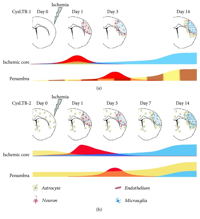
Figure 2.
Spatio-temporal expression of the CysLT1 and CysLT2 receptors after focal cerebral ischemia in rodents. (a) In the control brain, CysLT1 receptor is weakly expressed (time 0) [15, 61]. Following middle cerebral artery occlusion (MCAo), its expression, at the ischemic core level, is biphasic: at day 1 postischemia, the receptor is mainly expressed in neurons (red wave) [15, 60, 61] and, to a lesser extent, in astrocytes (orange) [15]; between 7 and 14 days postischemia, it increases in microglia (blue) [15]. In the boundary zone, that is, the “penumbra,” the receptor's expression is mainly expressed in neurons (red wave) at 3 days [60] and then it increases over time in most hypertrophic astrocytes (yellow) [15] and microvascular endothelial cells (brown) [15], reaching a peak after 14 days. (b) In the healthy brain, the CysLT2 receptor is primarily expressed in GFAP+ astrocytes around the lateral ventricles and in the cortex [18]. In the ischemic core, one day postischemia, the expression of CysLT2 receptor shows a rapid and transient peak in neurons (red) [18, 60] and then gradually disappeared over 3 days. In the hypertrophic microglia (blue), it slowly increases over time and reaches a peak after 14 days [18]. In the penumbra (boundary zone), following its induction at day 0, the receptor's expression is mainly expressed in neurons (red wave) at 3 days [60] and then it increases over time in astrocytes [18]. After one week, its expression also increases in the microglia [18].

