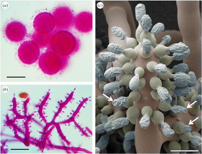
Figure 6.
Morphology of Escovopsis. Typical sporulating structures of Escovopsis taken directly from a cryptic midden sample (figure 1d), showing globose vesicles of (a) E. aspergilloides-type and cylindrical vesicles of (b) E. weberi-type, (c) CryoSEM of E. moelleri showing the large conidia produced on flask-shaped phialides from a clavate-cylindrical vesicle and developing distinctive wall ornamentation, apparently mucilaginous, with apical caps (arrows). Scale bars, (a,b) 30 µm, (c) 12 µm.

