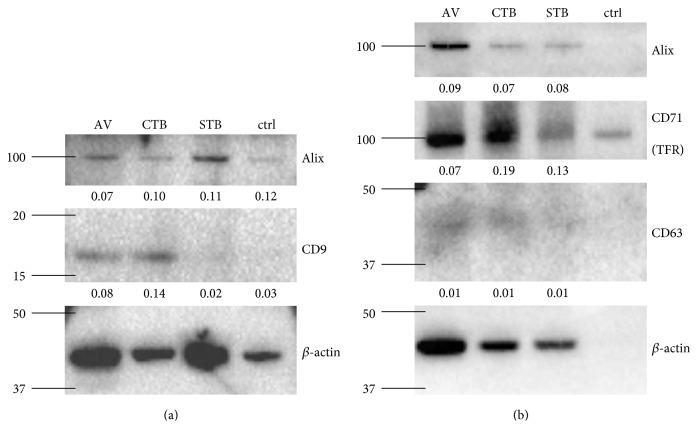
Figure 1.
EV isolation from ascites. EVs were isolated from ascites with the ligands AV (lane 1), CTB (lane 2), and STB (lane 3) and analyzed by western blotting. As a negative control, no ligand was added (lane 4). 250 μl of pooled ascites from 6 (a) or 16 (b) ovarian cancer patients were used as starting material. The tetraspanin proteins CD9 and CD63, the transferrin receptor (CD71), and the cytosolic protein Alix were examined, and β-actin was used as a loading control. The normalized band intensities of the analyzed proteins are given below each band. The molecular weight markers are indicated on the left side of the images in kDa.

