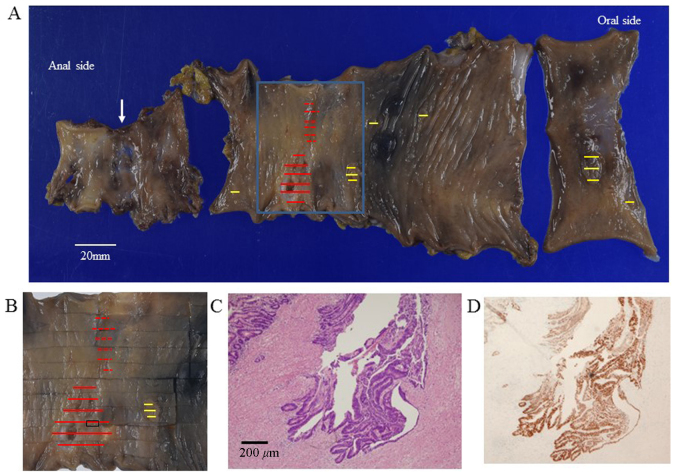
Figure 4.
Macroscopic findings of the resected specimen. (A) On macroscopic examination, the resected rectum and sigmoid colon contained two separate tumors and six flat polyps (yellow lines). The first tumor, invading the subserosa, was located 8 cm from the distal margin (red lines), and the second tumor, invading the submucosa, was located near the first (red dotted lines). The anastomosis site was located 4 cm from the distal margin (arrow). (B) Magnified image of the square indicated in (A). (C) On histopathological examination, the first tumor was diagnosed as well-differentiated adenocarcinoma with invasion of the subserosa [section as indicated in (B)]. (D) On immunohistochemical examination, p53 overexpression was observed in the first tumor.

