
Figure 1. How CTD phosphorylations modulate binding to Rtt103p CID .
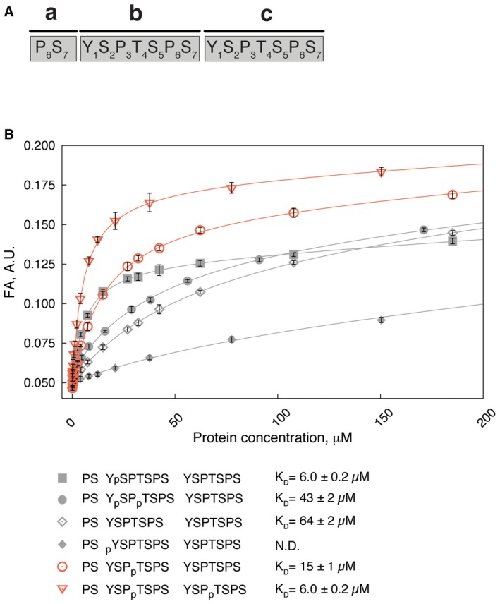
- Numbering of residues and order of heptad repeats of the CTD peptide used throughout the study.
- Equilibrium binding of Rtt103p CID with fluorescently labelled CTD peptides monitored by fluorescence anisotropy (FA). Rtt103p CID titrated into 10 nM FAM‐labelled CTD peptides. Peptide sequences, corresponding binding isotherms and dissociation constant (K D, ± standard deviation of the fit) are shown. FAM, 5,6‐carboxyfluorescein. N.D., not determined.