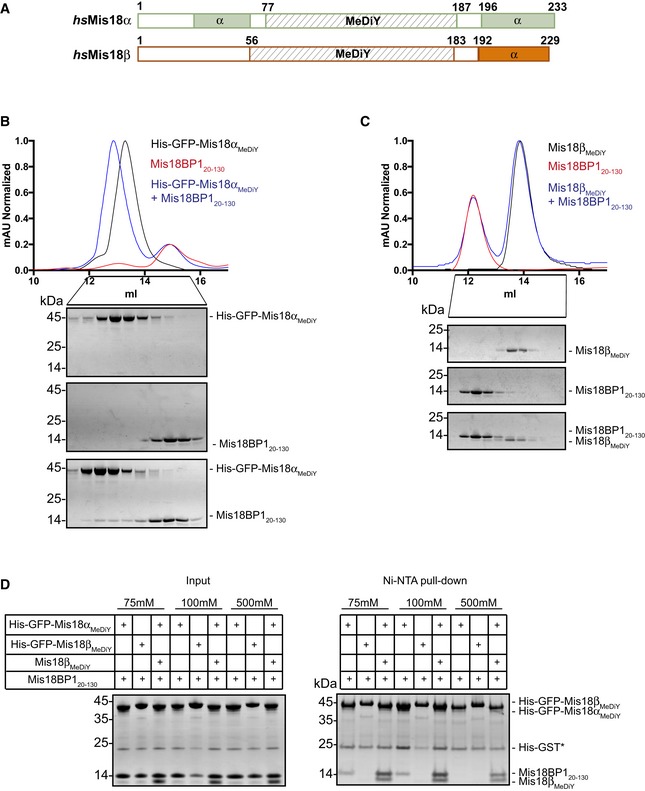
Figure 2. Mis18αMeDiY domain directly interacts with Mis18BP120–130 .

-
ASchematic representation of hsMis18α, hsMis18β domain architecture (using CDD and PsiPred).
-
B, CSEC profiles and respective SDS–PAGE analysis of (B) His‐GFP‐Mis18αMeDiY, Mis18BP120–130, and His‐GFP‐Mis18αMeDiY mixed with molar excess of Mis18BP120–130 and (C) Mis18βMeDiY, Mis18BP120–130, and Mis18βMeDiY with molar excess of Mis18BP120–130.
-
DSDS–PAGE analysis of the Ni‐NTA pull‐down assay where recombinant His‐GFP‐Mis18αMeDiY, His‐GFP‐Mis18βMeDiY, and Mis18βMeDiY were mixed with Mis18BP120–130 in different combinations in either 75, 100, or 500 mM NaCl. Left panel: inputs and right panel: Ni‐NTA pull‐down. * Contamination carried over from Mis18BP1 purification.
