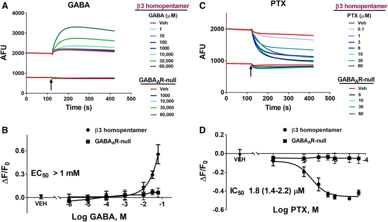
Fig. 5.
GABA potentiation and PTX inhibition of β3 homopentameric GABAAR-dependent FMP-Red-Dye fluorescence. HEK 293 cells transiently transfected with β3 homopentameric GABAAR were exposed to FMP-Red-Dye for 30 minutes to activate the receptors. GABAAR-null cells were used as the control. (A) GABA caused slow, concentration-dependent potentiation of the fluorescence in cells expressing β3 homopentameric GABAAR; minimal effects were obtained in GABAAR-null cells. (B) PTX caused slow, concentration-dependent inhibition of the fluorescence in cells expressing β3 homopentameric GABAAR; minimal effects were obtained in GABAAR-null cells. Arrows indicate time of addition of GABA and PTX, which was not removed. Red traces represent the responses to vehicle (0.01% dimethylsulfoxide). (B and D) Plots of ΔF/F0 from experiments similar to those illustrated in (A) and (C). Each data point represents mean ± S.D. of data from 10 wells. The EC50 value for GABA could not be determined since the response did not plateau. The IC50 value for PTX is 1.8 µM (95% confidence interval: 1.4–2.2 μM). Dose-response curves were plotted for PTX using nonlinear regression with a four-parameter logistic equation.

