Fig. 3.
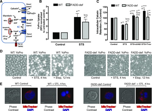
Caspase-3–cleaved Panx1 channels are functionally active in FADD-deficient Jurkat cancer cells during intrinsic apoptosis. (A) Schematic illustrates caspase-3–mediated proteolytic activation of Panx1 channel function as a pathway for ATP release or efflux/influx of charged organic dyes. (B, D) WT or FADD-deficient Jurkat cells were treated with 3 μM STS for 4 hours or 20 μM Etop for 12 hours. The cells were then washed, resuspended in BSS supplemented with 1 μM YO-PRO2+, and incubated for 20 minutes before quantification of YO-PRO2+ fluorescence per well or phase-contrast/epifluorescence imaging. (B) Data indicate mean ± S.E. from three independent experiments. Analysis by two-way analysis of variance and Tukey post-test comparison; a: +STS versus −STS; b: FADD-def versus WT. (D) Images are representative of two to three independent experiments with each stimulus. (C) WT or FADD-deficient Jurkat cells were loaded with 1 μM calcein-AM and then treated with STS for 4 hours in the presence or absence of 100 μM zVAD or 50 μM Trova. Cells were then washed and resuspended in BSS, and the calcein fluorescence was quantified. Data indicate mean ± S.E. from three independent experiments. Analysis by two-way analysis of variance and Tukey post-test comparison; a: +STS versus −STS; b: FADD-def versus WT; c: +STS with zVAD or Trova versus +STS alone. (E) WT or FADD-deficient Jurkat cells were treated with STS for 4 hours and then stained with MitoTracker Red and DAPI. Cells were imaged by confocal microscopy at original magnification 60×. Data are representative images of n = 15–19 individual cells. All panels: ns, not statistically significant; *P < 0.05; **P < 0.01; ***P < 0.001; ****P < 0.0001.

