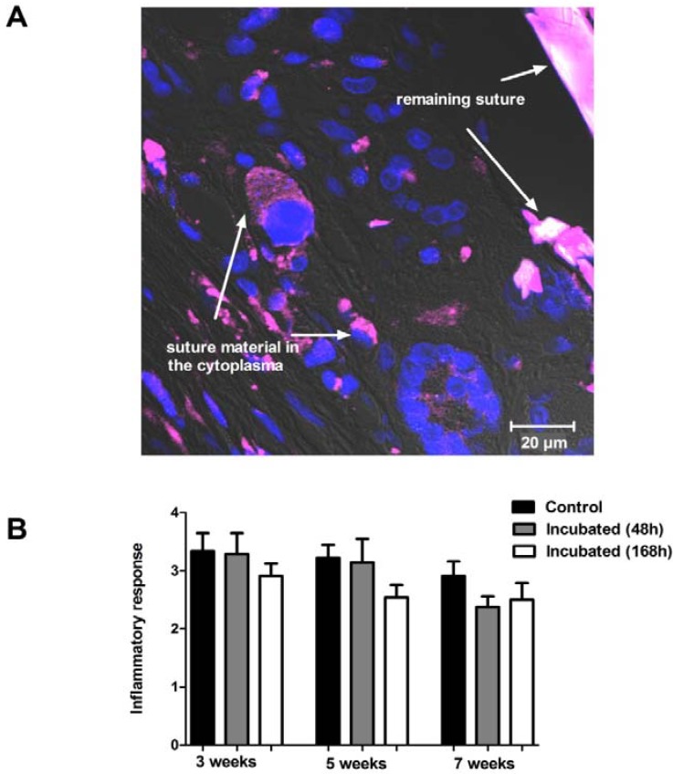
Figure 2.
Inflammatory response and phagocytosis of implanted suture material. Panel (A) shows a section from skeletal muscle with the remaining suture material (purple) 5 weeks after the implantation (63×). Blue color represents the nuclei of the cells. Degraded suture material can be seen in the cytoplasm of the surrounding cells. Panel (B) shows the inflammatory reaction to the control and to the 48 or 168 h pre-incubated suture material after 3, 5, or 7 weeks of implantation. No significant difference can be observed between the groups at any time point.

