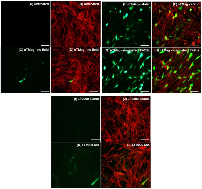
Figure 3.
Fluorescent microscopy images of NIH3T3 cells expressing GFP and correspondingly labeled with Phalloidin for actin stain of the whole cell population. (A,B) Untreated (C–L) transfected with 100 nm nTMag MNPs coated with pEGFPN1 plasmid DNA; in the absence of a magnetic field, for 30 min (C,D), in the presence of a static field (nanoTherics static array) for 30 min (E,F) and an oscillating field (nanoTherics magnefect-nano™ array at f = 2 Hz and amplitude = 200 µm), for 30 min (G,H), Lipofectamine 2000™ for 30 min (I,J) and Lipofectamine 2000™ for 6 h (K,L). Cell seeding density was 1 × 10⁴/96 well, incubation period (48 h, 37 °C, 5% CO2) post-transfection and scale bar = 100 μm in (A–L). GFP: green fluorescent protein; nTMag MNPs: nanoTherics nTMag magnetic nanoparticles; F: oscillation frequency.

