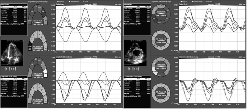
Figure 5.
(A): Four-chamber apical axis view from a patient with dilated cardiomyopathy. Please note that not only there is the presence of different temporal curves, suggestive of dyssynchrony; but also the opposite direction of curves, the latter showing abnormal relation at times of contraction that could be secondary to wall motion abnormalities. (B): Short axis view of the same patient. Please note the presence of different temporal curves, also suggestive of dyssynchrony.

