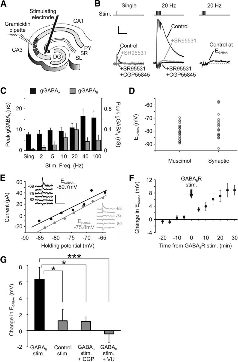
Figure 7.

Synaptically driven GABABR activation can shift EGABAA. A, Diagram of the experimental setup for synaptically activating postsynaptic GABAARs and GABABRs. Presynaptic GABAergic interneurons were stimulated in rat organotypic hippocampal slices via a bipolar tungsten electrode positioned at the stratum radiatum/pyramidale border, 50–100 μm from the recorded cell. B, Isolating GABAAR and GABABR responses. Representative traces show monosynaptic GABAergic postsynaptic currents in a CA3 pyramidal neuron recorded in response to single presynaptic stimuli (left) or trains of 6 stimuli applied at 20 Hz (middle). GABAAR and GABABR responses could be pharmacologically isolated by application of the selective GABAAR antagonist SR95531 (10 μm) and then the GABABR antagonist CGP55845 (5 μm). GABABR responses were not evoked by single stimuli but were evident for the multiple-stimuli condition. In the absence of these receptor blockers (right), the flux of chloride through GABAARs could be minimized by clamping the postsynaptic neuron close to its EGABAA. Calibration: 100 pA, 500 ms. C, The amplitude of the postsynaptic GABABR response is sensitive to presynaptic stimulus frequency. Whereas GABAAR conductances (gGABAA) were detected across the range of stimulus frequencies, GABABR-mediated conductances (gGABAB) were largest for high-frequency stimuli of ∼20 Hz and were minimal at lower frequencies (n = 9). D, Resting EGABAA values measured by muscimol activation of the GABAAR (n = 25) and by synaptic activation of the GABAAR (n = 22). Synaptic EGABAA exhibited a greater range of values and had a mean value of −76.7 ± 2.1 mV, compared with −81.1 ± 1.1 mV for the muscimol-evoked recordings (p = 0.06, t test). E, Example GABAAR I–V plots for a CA3 pyramidal neuron before (black data) and after (gray data) delivering a conditioning protocol designed to strongly activate postsynaptic GABABRs (90 stimuli delivered as 15 bursts of 6 stimuli at 20 Hz, at 5 s intervals). Insets, Raw traces. Calibration: 50 pA, 1 s. F, Change in EGABAA in a population of CA3 pyramidal neurons (n = 6) following delivery of the GABABR conditioning protocol (vertical arrow). G, CA3 pyramidal neurons that underwent the GABABR synaptic conditioning protocol (n = 6) showed a significantly larger positive shift in EGABAA than neurons that experienced a control stimulation protocol (90 stimuli delivered at 1 Hz) designed to generate minimal GABABR activation (n = 6, *p = 0.017, ANOVA followed by post hoc Dunnett's correction). The change in EGABAA induced by the GABABR synaptic conditioning protocol was also prevented by blocking GABABRs with the selective antagonist CGP55845 (n = 5, *p = 0.022) or by blocking KCC2 activity with VU0240551 (25 μm; n = 7, ***p = 0.001.
