
Figure 1.
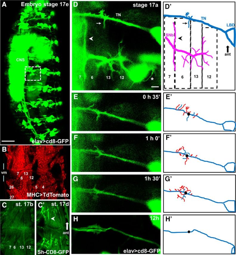
Live imaging of growth cones reveals that filopodia repeatedly contact off-target muscles during the last hours of embryogenesis. A, Pan-neural CD8-GFP expression in a Stage 17e Mhc1 mutant embryo (21 h AEL) shows unaffected nervous system development. The presence of all peripheral nerves, sensory cells, as well as a condensed CNS is evident. White square represents the area shown in D that includes the TN and the SNb nerve innervating the internal VL muscles 7, 6, 12, and 13 in the abdominal segment A3. Scale bar, 50 μm. B, Two adjacent hemisegments in an intact Mhc1 embryo expressing CD8-mCherry using the pan-muscular driver MHC-GAL4, revealing the presence of all body wall muscles. For orientation, the relative position of muscles 4, 5, 6, 7, 12, 13, 26, and 27 is labeled. C, The development of postsynaptic sites in Mhc1 embryos appears unaltered, as postsynaptic GFP-tagged potassium Shaker channels (MHC-Sh-CD8-GFP) transition from a dispersed (Stage 17b, 17 h AEL, C) to a restricted (Stage 17d, 20 h AEL, C′) localization in bouton-like structures (arrowhead) during embryogenesis. Muscles 6, 7, 12, and 13 are labeled for orientation. D, Motoneuron filopodia from growth cones on the TN and SNb nerve in a Stage 17a Mhc1 embryo (16.5 h AEL) as revealed by pan-neural GFP expression with a corresponding schematic showing the relative positions of muscles 6, 7, 12, and 13. Filopodial contacts simultaneously form on muscle 6 from correct (arrowhead) and incorrect (arrow) synaptic partners. Cell bodies from multiple sensory cells, including LBD that is part of the TN, are also fluorescent. The growth of sensory dendritic filopodia is evident (asterisk). Scale bar, 5 μm. E–H, Time-lapse snapshots of the TN in the same intact embryo at different time points, defining the image shown in D as the starting point. To illustrate filopodial dynamism, the corresponding schematics (E′–H′) depict filopodia extending off the TN at the time of the snapshot (blue) as well as filopodia that retracted 30 min before the snapshot (red). As reference, the black dot represents the origin of a prominent branch at time 0 shown in D.