
Figure 4.
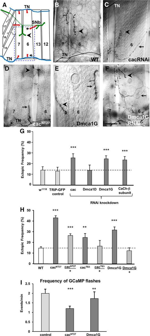
LOF of the voltage-gated Ca(v)2.1 cacophony and Ca(v)3 Dmca1G Ca channels leads to miswiring. A, Schematic of the musculature of the ventral portion of one abdominal hemisegment of a third instar larva showing muscle fibers 6, 7, 12, 13, 15, 16, and 17. On muscles 7 and 6, the ectopic contacts (red, arrows) are shown in the typical locations in which they are observed next to the native stereotypic motoneuron contacts (arrowhead). Blue represents TN. Green represents segmental nerve b (SNb). Gray represents segmental nerve d (SNd). Blue dashed square represents the area shown in images of ectopic contacts in Figures 4, 5, and 7. B–F, Ectopic contacts on muscles 7 and 6 were detected in third instar larvae in genetic manipulations that affect voltage-gated Ca2+ channels. Ectopic contacts comprised all different bouton types, ranging from ectopic Type 1b endings emerging from the TN (D) or from motoneurons innervating neighboring muscles (E), as well as Type II endings (C, F). C–F, Arrow indicates ectopic contact. Arrowhead indicates native innervation. Scale bars: B, D, F, 40 μm; C, 25 μm; E, 15 μm. G, Frequency of ectopic contacts in larvae expressing RNAi constructs for all genes encoding for α subunits of voltage-gated Ca2+ channels, cacophony (cac, Ca(v)2.1), Dmca1D (Ca(v)1), and Dmca1G (Ca(v)3), as well as for the Ca2+ channel β subunit, using the pan-neural elavGAL4C155 driver. Control groups included elavGAL4 flies crossed to either W1118 or UAS-GFP flies, and experimental lines were compared with their corresponding control for statistical significance. Data are calculated as the percentage of ectopic-containing hemisegments per larva, averaged over the number of animals (±SEM; n = 19, 12, 22, 8, 26, and 18 animals, respectively; p = 0.005, 0.006, and 0.008, respectively). H, Frequency of ectopic contacts in WT and homozygous and heterozygous larvae for cacNT27, cacTS3, and Dmca1G (n = 51, 36, 61, 17, 10, 34, and 18 animals, respectively; p = 1.1 × 10⁻¹⁸, 8.8 × 10⁻⁸, 0.01, and 1.1 × 10⁻⁶, respectively). I, Frequency of GCaMP5 flashes in Stage 17e embryos in control, cacNT27, and Dmca1G homozygotes, measured at a single motoneuron as described in Figure 3 (n = 21, 13, and 13, respectively; p = 0.0004 and 0.009, respectively).