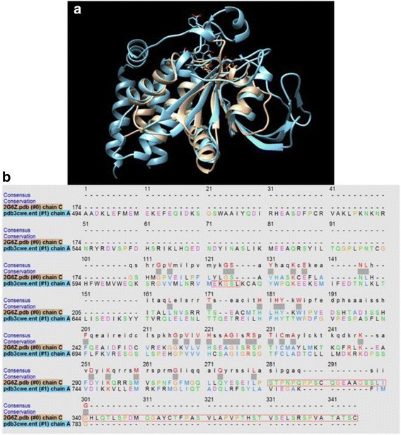
Fig. 9.

Comparison of PTP1B and DUSP5 PD. a Structural overlay of PTP1B and DUSP5 PD(WT), based on crystal structures with pdb codes 3CWE (PTP1B with a phosphonic acid inhibitor bound) and 2G6Z (DUSP5 PD), using Chimera. b Primary sequence alignment was done using Clustal Omega pairwise alignment and guide tree algorithm [37], which indicated an extremely low percent identity of 9.5%
