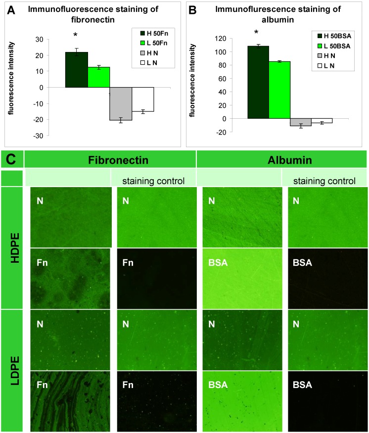
Figure 2.
Immunofluorescence staining of (A) fibronectin; and (B) albumin and their microphotographs (C) on HDPE (H) and LDPE (L), in non-treated form (N), treated with plasma for 50 s and subsequently grafted with fibronectin (50 Fn) or bovine serum albumin (50 BSA). Olympus IX 51 microscope, obj. 10x, DP 70 digital camera. The measurements were performed at the same exposure time for all experimental groups (1.2 s).

