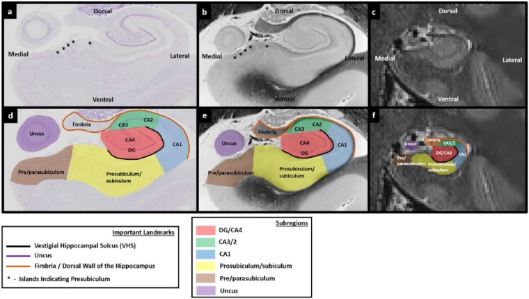
Figure 1.
Subregions of the human hippocampus. (a) A section of post-mortem human hippocampus stained with cresyl violet to visualise cell bodies and (b) an equivalent slice of post-mortem human hippocampus stained with haematoxylin (Weigert) to visualise white matter. Both sections are from ‘The Human Brain’ website http://www.thehumanbrain.info/brain/sections.php. (c) A T2-weighted structural MRI of the human hippocampus. This section is approximately equivalent to the slice represented in ‘a’ and ‘b’. (d) The same section is presented as in ‘a’ but now overlaid with hippocampal subregion masks. (e) The same section is presented as in ‘b’ but now overlaid with hippocampal subregion masks. (f) The same section is presented as in ‘c’ but now overlaid with hippocampal subregion masks.

