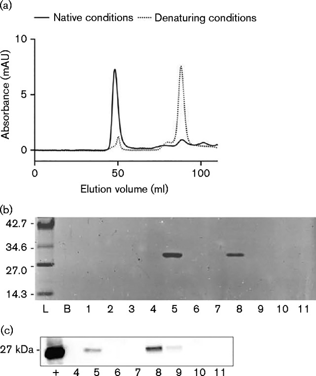
Fig. 4.

Determining the oligomeric state of purified His6-PspA protein. (a) Size exclusion chromatography trace of His6-PspA showing a void volume at 49.5 ml containing the aggregated and higher-order PspA protein species, and a collection of peaks between 83.0 and 109.0 ml containing putative dimeric and monomeric His6-PspA species. Included is an overlay of His6-PspA protein purified under denaturing conditions. AU, absorbance units. (b) SDS-PAGE of His6-PspA purified by gel filtration. Lanes represent pooled fractions from 0–10 (lane 1), 10–20 (lane 2), 20–30 (lane 3), 30–40 (lane 4), 40–50 (lane 5), 50–65 (lane 6), 65–80 (lane 7), 80–90 (lane 8), 90–100 (lane 9), 100–110 (lane 10) and 110–120 ml (lane 11). Values in kDa. (c) Western blot confirming the presence of His6-PspA protein. Fractions are the same as those outlined in (b);+, His6-PspA positive control.
