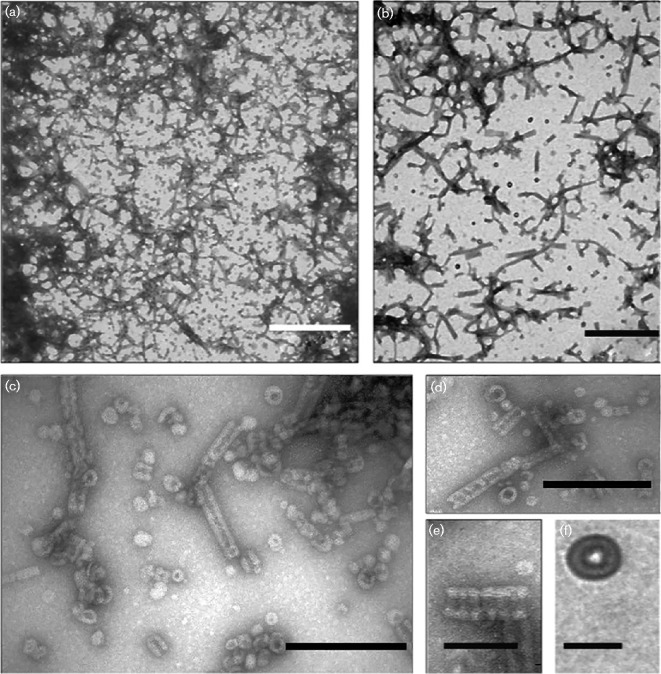
Fig. 5.
Transmission electron micrographs of negatively stained PspA protein complexes. (a, b) Mixtures of rings, rod-like complexes and mesh-like structures are readily visible. Bar, 500 nm. (c, d) Both the ring structures and rod-like complexes are visible in this field of view. Bar, 200 nm. (e) Close-up of a rod-like structure, clearly showing the indentations and striations that indicate stacking of ring-like structures. This is also an example of the tapered end observed occasionally. Bar, 100 nm. (f) The putative 36-mer, ring-like PspA structure. Bar, 40 nm.

