FIG 2.
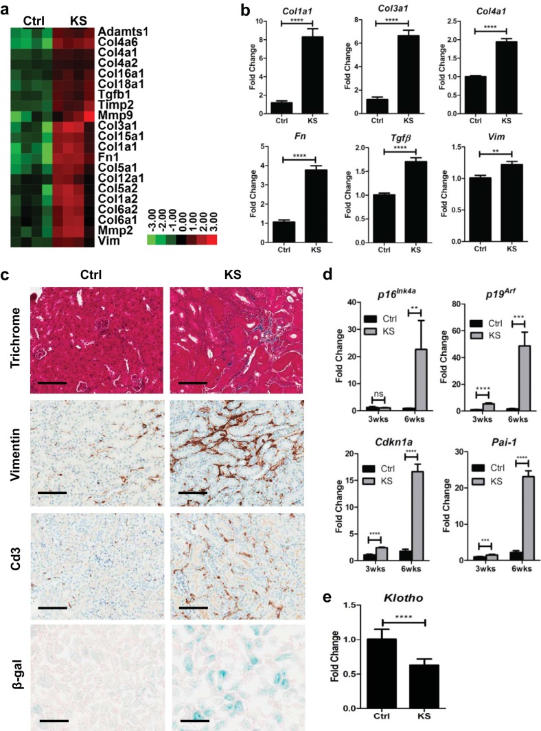
Sav1 deletion in renal tubule cells activates profibrotic and cell senescence genes. (a) Heat map representation showing increased expression of genes associated with renal fibrosis in renal cortex tissue from KspCreER; Sav1f/f mice 6.7 weeks after treatment with vehicle (Ctrl) or tamoxifen (KS). (b) Real-time qRT-PCR analysis of total RNA isolated from renal cortex tissue of KspCreER; Sav1f/f mice 3 weeks after treatment with vehicle (Ctrl; n = 4) or tamoxifen (KS; n = 4). (c) Representative photomicrographs showing Ctrl (n = 6) and KS (n = 6) kidney sections 6 weeks after treatment, stained with Masson's trichrome and stained for vimentin, Cd3, and senescence-associated β-galactosidase (SA-β-Gal). (d) Relative expression of senescence genes assessed after treatment with vehicle (Ctrl; n = 4) or tamoxifen (KS; n = 4) for the indicated times. (e) Relative Klotho gene expression in Ctrl (n = 4) and KS (n = 4) mice as assessed by real-time qRT-PCR 3 weeks after treatment with vehicle or tamoxifen. All relative expression values from real-time qRT-PCR analyses were normalized to the 18S rRNA gene expression level. All vehicle-treated mice were given sunflower oil by oral gavage. Error bars in all graphs represent standard deviations. ns, not statistically significant; **, P ≤ 0.01; ***, P ≤ 0.001; ****, P ≤ 0.0001. Magnification, ×20 for all images; bars = 100 μm.

