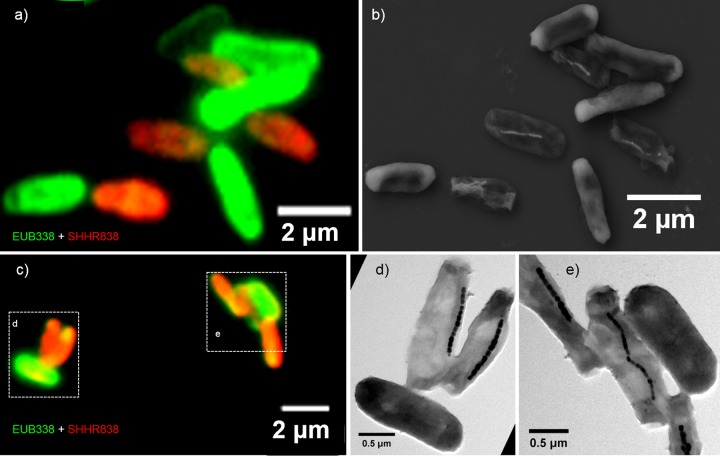
FIG 3.
Fluorescence-coupled electron microscopy identification of SHHR-1 cells. (a) Overlapping fluorescence microscopy image of SHHR-1 and E. coli cells mounted on the surface of a cover slide glass and in situ hybridized with the 5′-FAM-labeled universal bacterial probe EUB338 (green) and the 5′-Cy3-labeled SHHR838 probe (red). (b) Coordinated SEM image of the same field as in panel a. (c) Overlapping fluorescence microscopy image of SHHR-1 and E. coli cells mounted on the surface of a TEM grid and in situ hybridized with the 5′-FAM-labeled universal bacterial probe EUB338 (green) and the 5′-Cy3-labeled SHHR838 probe (red). (d) Coordinated TEM image of the same field indicated by dashed-line box (left) as in panel c. (e) Coordinated TEM image of the same field indicated by dashed-line box (right) as in panel c. Those bacteria that are only fluorescently labeled with the EUB338 probe and do not contain magnetosomes are inner-control E. coli cells. In contrast, those bacteria that are fluorescently labeled with both the EUB338 and SHHR838 probes (yellow-red colors) and contain magnetosomes are SHHR-1 cells.

