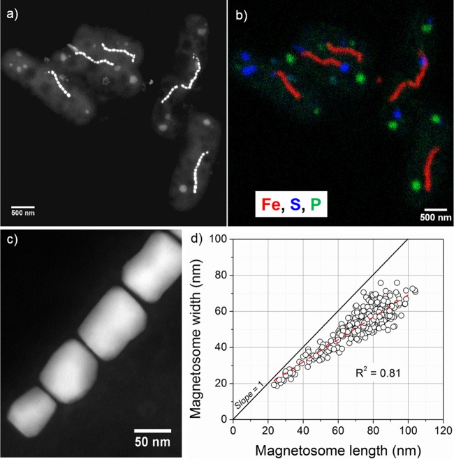
FIG 4.
Morphological and chemical features of SHHR-1 cells. (a) HAADF-STEM image of five SHHR-1 cells. (b) Chemical composition map of the same five SHHR-1 cells as shown in panel a. HAADF-STEM imaging and STEM-EDXS mapping analyses show that SHHR-1 cells contain magnetite-type magnetosomes as single chains, as well as irregular polyphosphate and sulfur-rich inclusions. (c) High-magnification HAADF-STEM image of SHHR-1 magnetosomes showing their prismatic shapes and chain alignment along the long axes of individual particles. (d) Plot of crystal length versus width showing a linear relationship between crystal length and width of SHHR-1 magnetosomes.

