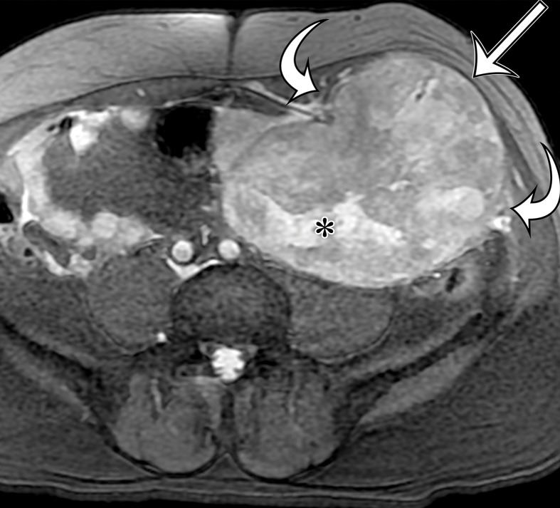
Figure 10d.
Malignant perivascular epithelioid cell tumor in a 56-year-old man with crampy left lower quadrant pain. (a, b) Axial (a) and sagittal (b) contrast-enhanced CT images show a heterogeneously enhancing soft-tissue mass inseparable from the anterior abdominal wall musculature (straight arrow), with central low-attenuation areas of necrosis (* on a) and punctate calcifications (curved arrow). (c, d) Axial T2-weighted fat-suppressed (c) and contrast-enhanced T1-weighted (d) MR images show heterogeneous T2 hyperintensity and avid enhancement within the cellular portions (straight arrow), with more focal hyperintensity and lack of enhancement of the necrotic portions (*). Abdominal wall invasion (curved arrows) is more apparent on the MR images. (e) Photograph of the cut surface of the resected specimen shows corresponding areas of necrosis and hemorrhage (*) within the predominantly solid lobulated mass. Mass is inseparable from the abdominal wall (arrow).

