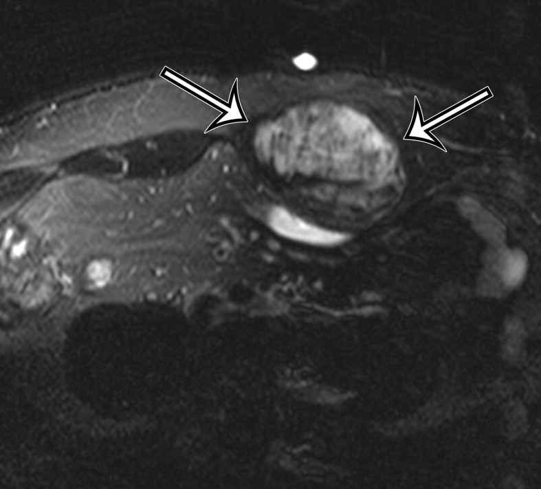
Figure 11b.
Desmoid tumor incidentally identified in the anterior abdominal wall of a 43-year-old man. (a) Axial contrast-enhanced CT image shows heterogeneous expansion of the left rectus muscle, with a suspected mass in the medial aspect of the muscle (arrows). (b, c) Axial T2-weighted fat-suppressed (b) and contrast-enhanced T1-weighted (c) MR images obtained to better characterize the lesion show a heterogeneous predominantly hyperintense solid mass (arrows on b) with heterogeneous enhancement (arrows on c).

