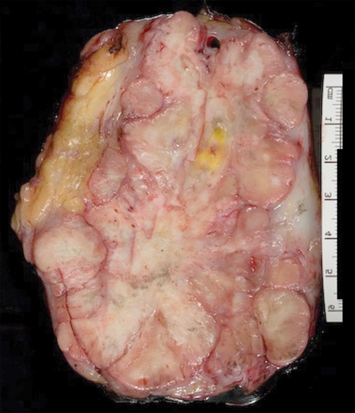
Figure 4c.
Solitary fibrous tumor in a 69-year-old man with long-term lower urinary tract symptoms and back pain radiating to the right leg. (a, b) Axial (a) and coronal (b) contrast-enhanced CT images show marked heterogeneous enhancement of a sharply marginated lobulated solid mass (straight arrow) in the right pelvis. A prominent peripheral blood vessel (curved arrow) drapes over the superior margin of the mass. (c) Photograph of the cut surface of the resected specimen shows the encapsulated pink-tan soft-tissue mass. (Scale is in centimeters.)

