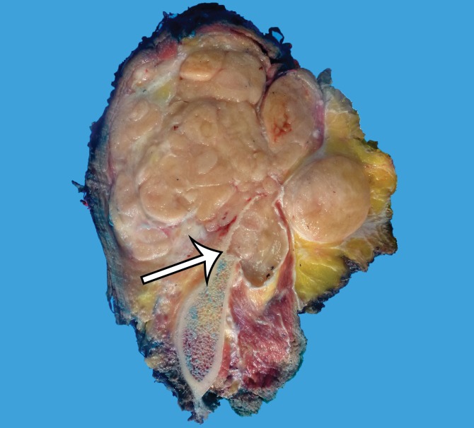
Figure 5d.
MPNST in a 34-year-old woman with low back pain radiating to the left lower extremity. (a–c) Coronal T2-weighted inversion-recovery (a), T1-weighted (b), and contrast-enhanced T1-weighted fat-suppressed (c) MR images show a heterogeneous lobulated left paraspinal mass (arrow) invading the L5 vertebral body medially, the sacrum inferomedially, and the iliac wing inferolaterally. The mass is T1 hypo- to isointense (arrow on b) and T2 hyperintense (arrow on a) relative to skeletal muscle, with avid heterogeneous enhancement (arrow on c). (d) Photograph of the cut surface of the resected specimen shows a multinodular pale yellow–tan mass invading the iliac wing (arrow).

