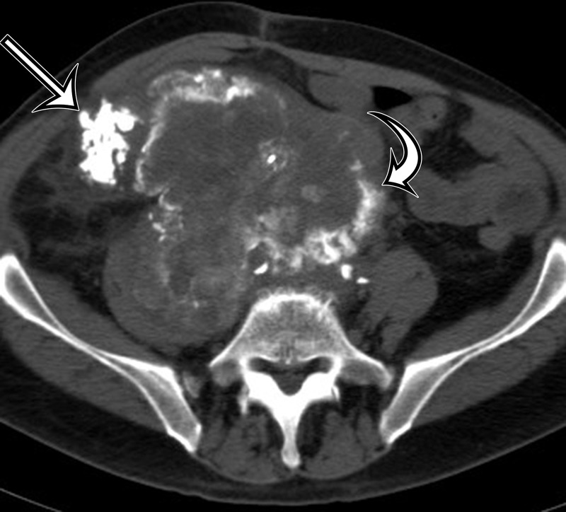
Figure 6a.
Soft-tissue osteosarcoma in a 59-year-old man with right flank pain. (a) Axial CT image (bone window) shows dense calcification (straight arrow) and an osteoid matrix (curved arrow) in a lobulated right pelvic mass. (b) Axial T2-weighted fat-suppressed MR image shows central areas of markedly T2-hyperintense necrosis (*). (c) Axial contrast-enhanced T1-weighted fat-suppressed MR image shows enhancement of the periphery (arrow) of the mass.

