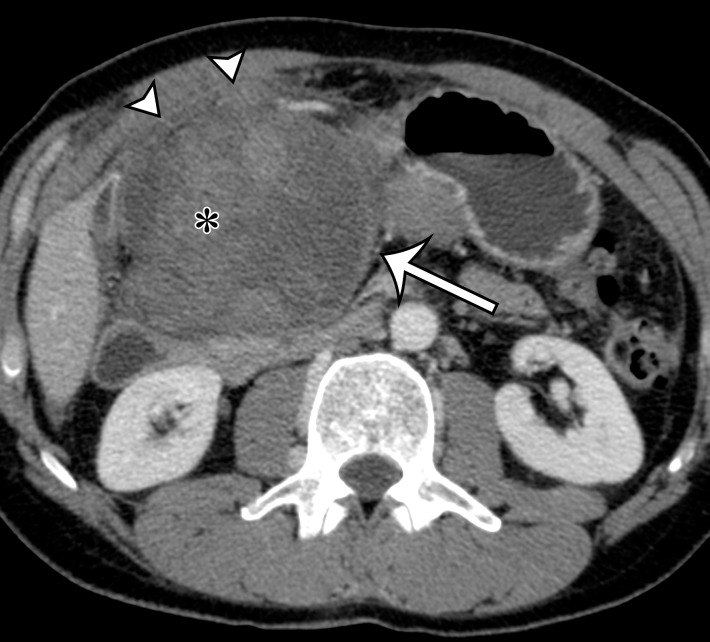
Figure 8c.
Synovial sarcoma in two different patients. (a) Synovial sarcoma in a 59-year-old woman who presented with rapid enlargement of a palpable painless mass: Axial contrast-enhanced CT image shows a well-circumscribed heterogeneously enhancing mass within the right anterior abdominal wall (arrows), with cystic components (*). (b, c) Synovial sarcoma in a previously healthy 36-year-old man with new onset of intractable vomiting: Axial contrast-enhanced CT images (c obtained at a lower level than b) show a large heterogeneous mass (straight arrow) infiltrating along the gastrohepatic ligament and circumferentially invading the gastric antrum and lesser curvature (curved arrows on b), with mass effect on the liver, gallbladder, duodenum, and pancreas. Areas of nonenhancing hyperattenuation are consistent with intratumoral (*) and intraperitoneal (arrowheads on c) hemorrhage.

