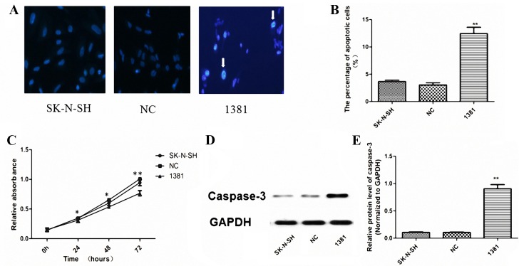
Figure 3.
Apoptosis, proliferation and caspase-3 activation in 1381 cells. (A) Hoechst 33342 nucleic acid staining in untransfected SK-N-SH, NC-transfected and 1381 cells. Arrows indicate the characteristic appearance of nuclear chromatin condensation and nuclear fragmentation in apoptotic 1381 cells. Magnification, ×400. (B) The percentage of apoptotic cells in the 1381 group was significantly increased compared with that in NC and untransfected SK-N-SH cells (**P<0.01). (C) CCK-8 assay demonstrated that the miR-21-inhibitor significantly reduced the proliferation of 1381 cells compared with the viability of SK-N-SH and NC cells at 24, 48 and 72 h. WB analysis illustrating a (D) representative image from triplicate experiments and (E) quantitative results, indicating a significantly increased level of activated caspase-3 protein when miR-21 was inhibited in 1381 cells, compared with that in NC and SK-N-SH cells. *P<0.05, **P<0.01 vs. NC and SK-N-SH. NC, negative control; miR, microRNA; 1381, SK-N-SH cells transfected with miR-21 inhibitor; WB, western blot; CCK, cell counting kit.

