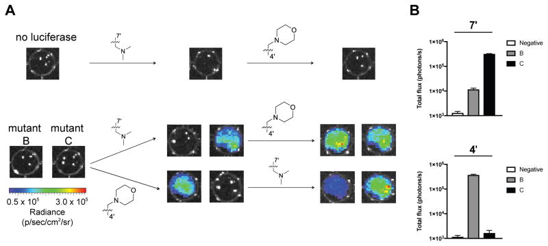
Figure 5. Imaging cells with orthogonal luciferase-luciferin pairs.
(A) Mutant luciferase-expressing DB7 cells were plated (1.5 x 10⁵ cells/well) in 96-well black plates and sequentially incubated with C4′ and C7′ sterically modified luciferins (750 μM). Representative bioluminescence images are shown. (B) Quantification of the images from (A) after initial substrate addition. Error bars represent the standard error of the mean for experiments performed in triplicate.

