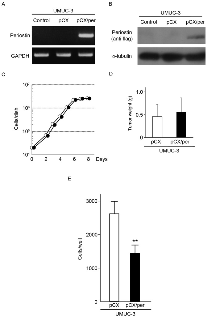
Figure 1.
Effect of ectopic expression of periostin on cell invasiveness, cellular proliferation and subcutaneous tumor growth in UMUC-3 cells. (A) RT-PCR analysis of periostin in UMUC-3 cells infected with pCX or pCX/Per. GAPDH expression was used as an internal control. (B) Immunoblot analysis of exogenous periostin in UMUC-3 cells infected with pCX or pCX/Per. Exogenous expression of periostin was detected by anti-Flag antibody. α-tubulin was used as an internal control. (C) Growth curves of UMUC-3 cells infected with vector virus or periostin-expressing virus. Experiments were performed in triplicate. SDs were too small to show using error bars. (D) Subcutaneous tumor growth of UMUC-3 cells infected with vector virus or periostin-expressing virus. Bar ± SD of three mice for each cell line. P=0.691. (E) Suppression of cell invasiveness of UMUC-3 cells by periostin expression. Number of cells invading through the Matrigel is shown. Each sample was assayed in triplicate. Bar ± SD of triplicate chambers for each experiment. **P=0.006 vs. pCX. pCX, control virus; pCX/Per, periostin-expressing virus; white circle, vector virus; black circle, periostin-expressing virus; SD, standard deviation; white bar, vector virus; black bar, periostin-expressing virus.

