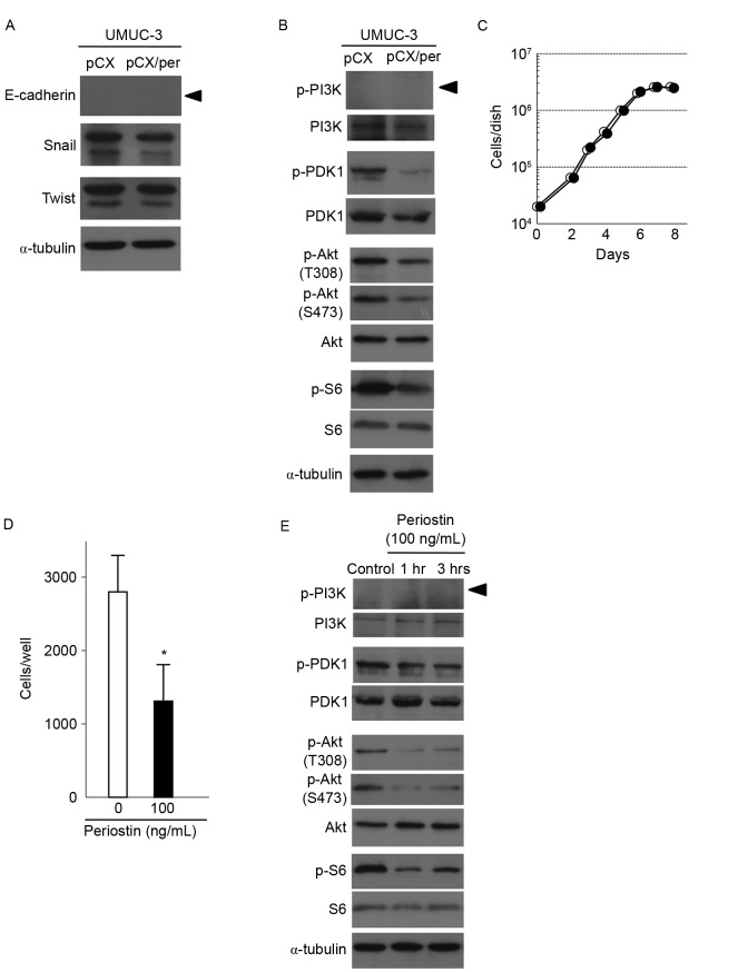
Figure 3.
Effects of the expression of periostin on PDK1/Akt/mTOR signaling and expression of proteins regulating epithelial-mesenchymal transition in UMUC-3 cells. (A) Immunoblot analyses of the EMT-associated proteins E-cadherin, Snail and Twist in vector control and periostin-expressing UMUC-3 cells. α-tubulin expression was used as an internal control. The arrowhead indicates the E-cadherin band. (B) Immunoblot analyses of total and phosphorylated forms of PDK1, Akt and S6, a downstream protein of mTOR, in vector control and periostin-expressing UMUC-3 cells. α-tubulin expression was used as an internal control. The arrowhead indicates the p-PI3K band. (C) Growth curves of UMUC-3 cells treated with 100 ng/ml periostin and vector control cells. Experiments were performed in triplicate. SD was too small to show as error bars. White and black circles indicate the control and periostin treatment (100 ng/ml), respectively. (D) Suppression of cell invasiveness of UMUC-3 cells following treatment with 100 ng/ml periostin for 6 h. Number of cells invading through the Matrigel is shown. Bar, ± SD of triplicate chambers for each experiment. *P=0.046 vs. control cells. (E) Immunoblot analyses of total and phosphorylated forms of PDK1, Akt and S6 in control UMUC-3 cells and cells treated with 100 ng/ml periostin for 1 and 3 h. α-tubulin expression was used as an internal control. The arrowhead indicated the p-PI3K band. PDK1, phosphoinositide-dependent kinase-1; Akt, protein kinase B; mTOR, mammalian target of rapamycin; SD, standard deviation.

