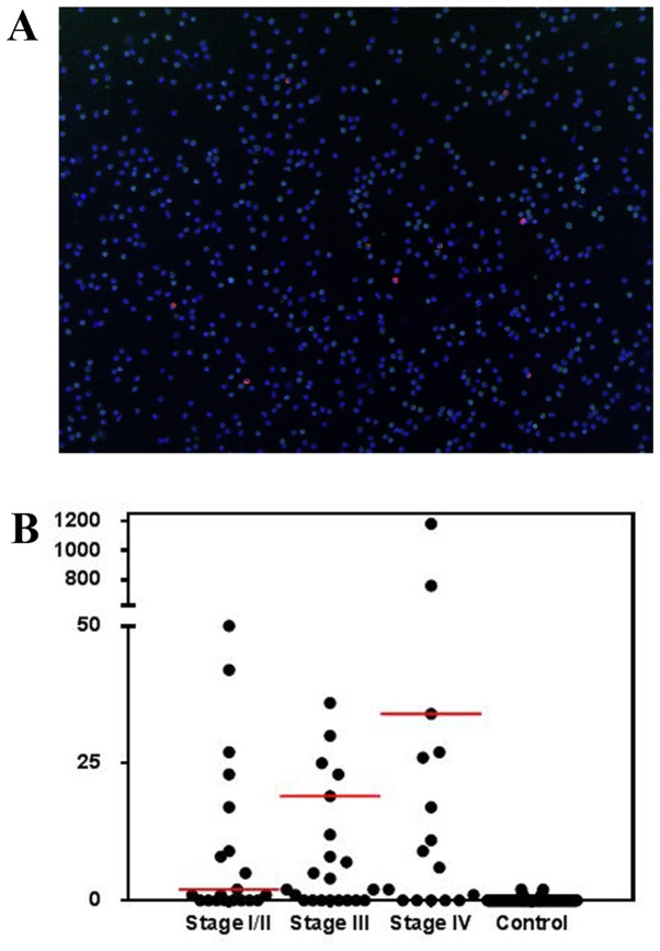
Figure 2.
Samples from patients with various stages of lung cancer were examined using the unbiased detection method. (A) A typical image of CTCs detected by a high-content imaging system (version 10.8; IN CELL Analyzer). Magnification, ×4. Green light, anti-cluster of differentiation 45; red light, anti-pan cytokeratin; blue, DAPI. (B) The distribution of CTC counts in patients with lung cancer according to tumor stage. The red lines illustrate the median. CTC, circulating tumor cell.

