Figure 1.
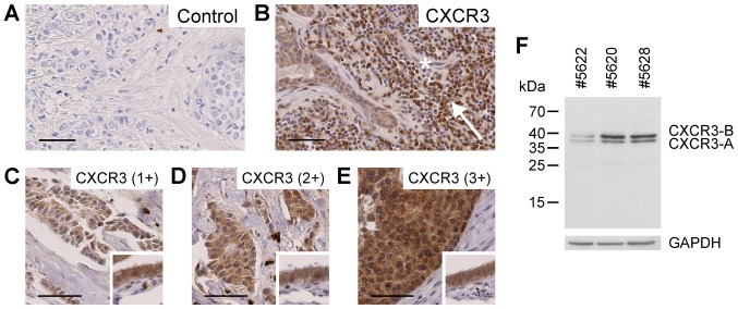
Protein expression of CXCR3 in human invasive ductal breast cancer formalin-fixed paraffin-embedded specimens, as evaluated by immunohistochemistry. (A) Negative control without addition of primary antibody. (B) Expression of CXCR3 in tumor-adjacent normal ductal cells, tumor-infiltrating lymphocytes (arrow) and endothelial cells (asterisk). (C-E) Various breast cancer tissues with increasing CXCR3 protein expression. Small inserts include fallopian tube epithelium as a positive control for CXCR3 protein expression. (F) Specific detection of the CXCR3 splice variants CXCR3-A and CXCR3-B by western blot analysis using MAB160 antibody in three human breast cancer tissue extracts. Scale bars, 50 µm. CXCR3, C-X-C motif chemokine receptor 3.

