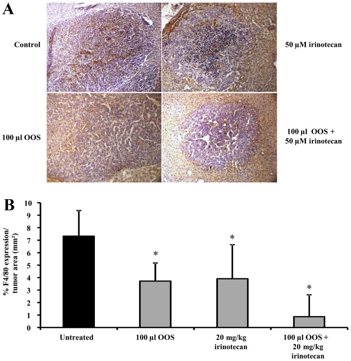
Figure 5.
Effect of the combined treatment of irinotecan and OOS® on the tumor infiltration of macrophages in vivo. Expression level of F4/80 was analyzed in liver tissue by immunohistochemistry. (A) Images showing F4/80 expression (brown) and hematoxylin (purple) in liver tissue collected from untreated and treated mice. Image magnification was ×20. (B) F4/80 expression was quantified in livers collected from untreated and treated C26-bearing mice. Data are calculated as % of F4/80 expression per tumor foci area. At least 6 mice per group were used and differences were considered statistically significant at *P<0.05.

