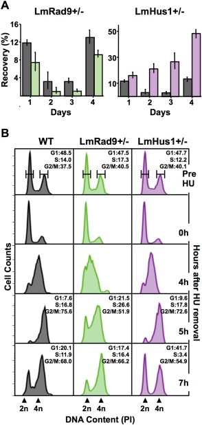
Figure 3.

LmRad9 and LmHus1 have distinct roles in the response to replication stress.
A. WT (gray), LmRad9+/− (green) and LmHus1+/− (purple) cells were treated with 10 mM HU for ∼15 h and seeded in drug‐free media at 105 cells/ml; cell densities were assessed daily and recovery was calculated as a percentage of proliferation as compared with the nontreated cells; vertical lines on top of each bar indicate standard deviation.
B. Cell cycle progression analysis of WT, LmRad9+/− and LmHus1+/− cell lines; cell cycle were blocked with 5 mM HU for 8 h, seeded in HU‐free medium and collected at the indicated time points; DNA content was examined by flow cytometry; each histogram represent data from 10,000 events; 2n and 4n indicate nonreplicated and replicated DNA, respectively; percentage of cells in gated G1, S, and G2/M phases is indicated for cells before HU treatment (Pre HU) and for cells at 5 and 7 h after HU removal.
