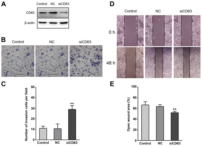
Figure 4.
Effect of CD63 on esophageal cancer cell invasiveness. (A) Western blot analysis confirmed a marked reduction of CD63 protein following CD63 small interfering RNA transfection in esophageal cancer cells. (B and C) In the Matrigel invasion assay, following treatment for 48 h, the number of CD63 knockdown TE-1 cells that migrated to the bottom surface of the membrane was greater compared with TE-1 cells transfected with empty plasmid and the control TE-1 cells. (D and E) Wound healing was observed 48 h after treatment, and the open wound area of CD63 knockdown TE-1 cells was larger than the TE-1 cells transfected with empty plasmid and the control TE-1 cells. **P<0.01. NC, negative control; siCD63, small interfering RNA targeting CD63.

