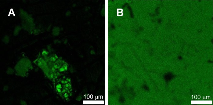
Figure 1.
Laser scanning confocal microscope images (0.5 × 0.5 mm) of films obtained by blending water dispersions of fluorescent SiNP with poly(butyl methacrylate) (PBMA) nanoparticles of a 100-nm diameter using the same silica volume fraction as in the core-shell particles show aggregates of fluorescent silica particles and large dark polymer domains (A). Films cast from core-shell water dispersions show a homogeneous fluorescence distribution (B). Both films were cast from a water dispersion, dried at 32 °C, and annealed at 90 °C for 1 h. Reprinted with permission from [9]. Copyright 2009, American Chemical Society.

