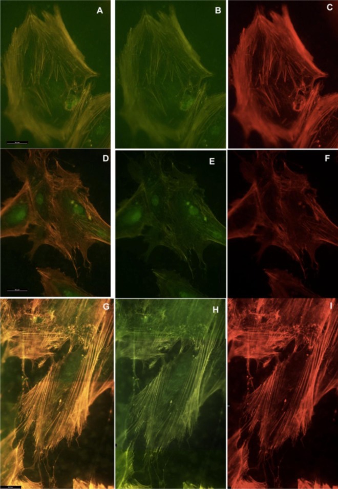
Figure 5.
HOB cells grown on TiO2 functionalized membranes, and imunolabeled with rhodamine-phalloidine for actin cytoskeleton (red) and antivinculin antibody (green) for focal adhesion sites, 72 h after seeding. Representative images of cytoskeletal features, showing the differences in stress fibers, and focal adhesion sites vinculin-dependent. Osteoblasts grown on (A,B,C) TiO2/PLGA-10; (D,E,F) TiO2/PLGA-30; (G,H,I) TiO2/PLGA-100. Combined, overlay, images for both rhodamine-phalloidine labeling and antivinculin antibody for each group are shown in (A,D,G). Magnification 40×. Scale bar = 50 μm.

