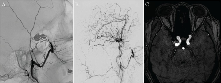
Fig. 3.
(A) The right external carotid angiogram after sinus embolization (lateral) shows the coil mass in the isolated cavernous sinus and the feeding arteries. (B) The right common carotid angiogram after treatment (lateral) shows total occlusion of the fistula. (C) The postoperative time-of-flight magnetic resonance image confirms the disappearance of the shunt.

