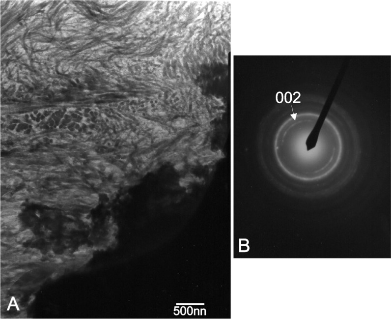
Figure 10.

(A) Transmission electron micrograph of the stained interface at the 15th month, showing well resolved collagen fibers in the new formed Ca-P phase; (B) Equivalent selected area diffraction pattern of the new bone tissue; unstained thin section.
