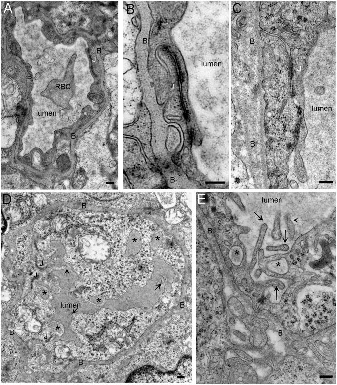
Figure 2.
Morphology of the capillaries is modified in the tumor but tight junctions are still intact. (a) Transverse section of control capillary. The capillary wall is formed by thin endothelial cells that are connected by tight junctions (J). Surrounding the capillary is a basal lamina (B) beyond which is a covering of astrocytic processes. (b, c) Junctions between endothelial cells in control (b) and tumor capillaries (c). No difference in the length or the morphology is observed between the tight junctions of control and tumor capillaries. (d) Tumor capillary. Several membrane ruffles protrude in the lumen of the capillaries and the shape as well as the structure of the basal lamina is irregular. There are numerous macropinosomes (*) inside the endothelial cell. The electron density of these compartments is similar to that of the capillary lumen. (e) Detail of an endothelial cell. Membrane protrusions in the capillary lumen form cup shape profile that sometime fuse together to form macropinosome (*). Notice the disorganization of the basal lamina (c, d, and e). RBC: red blood cell (bar: 200 nm).

