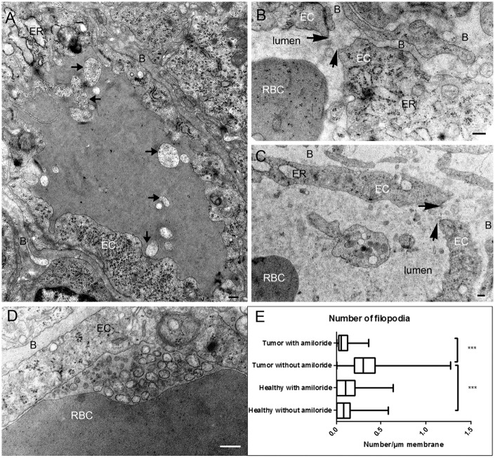
Figure 3.
Tumor capillaries of amiloride-treated rats. (a) Cross section of a capillary. Dilatation of the endoplasmic reticulum (ER) could be observed in the endothelial cell, and heterogeneous membranes are present in the lumen of the capillary (arrows). (b, c) Opening of the junction (arrows) observed in amiloride-treated rats. Components of the blood diffuse in the basal lamina. (d) Accumulation of membranes vesicles in the lumen of the capillary. (e) Quantification of the number of protrusion in the lumen divided by the circumference of the capillary. Data are presented as boxplots with min/max values. B: basal lamina; EC: endothelial cell; ER: endoplasmic reticulum; RBC: red blood cell (bar: 200 nm).

