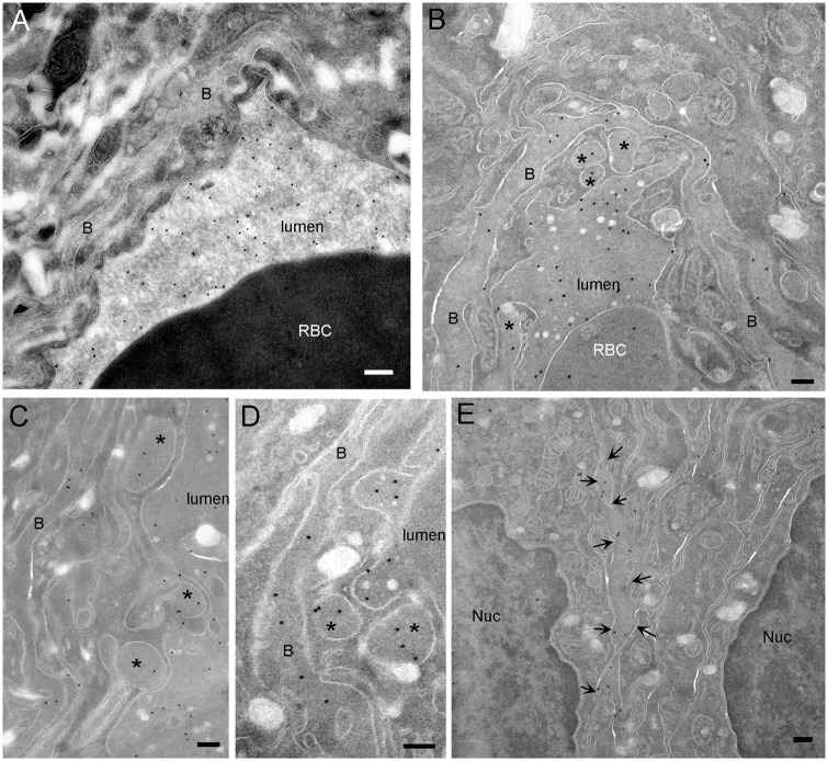
Figure 4.
IgG diffuse from the lumen of tumor blood vessel to the extracellular space. (a) Control capillary. Protein A-gold labeling on cryosection shows that endogenous IgGs are located in the lumen of capillaries. Gold labeling is neither observed in the endothelial cells nor in the basal lamina. (b) In tumor, IgGs are located in the lumen of the capillaries, in the basal lamina and in macropinosomes (*). (c, d) In tumor, IgGs are located in macropinosomes inside the endothelial cell as well as in the basal lamina. Notice that IgG does not contact macropinosome membranes. (e) In tumor, protein A-gold labeling of IgG shows that endogenous antibodies diffuse between the tumor cells (arrows). B: basal lamina; Nuc: nucleus; RBC: red blood cell (bar: 200 nm).

