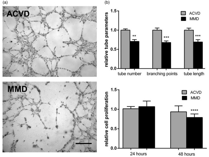
Figure 4.
Impaired cEND tube forming activity and cell proliferation in response to MMD serum. (a) CEND cell proliferation measured at 24 or 48 h of ACVD and MMD serum incubation determined employing MTT proliferation assay and expressed as cell proliferation rate relative to the ACVD control. (b) Phenotypic effect of ACVD and MMD serum incubation tested by tube formation assay. Representative low magnification images (10 × ) of tubes formed by serum-conditioned cENDs were chosen. Total tube number, branching points, and tube length were assessed by Wimasis Software. Values are means ± SEM, (n = 3 independent experiments each including five different patients’ sera per group performed in triplicates), ****p < 0.001, ***p < 0.001, **p < 0.01 versus cENDs incubated in ACVD serum.

