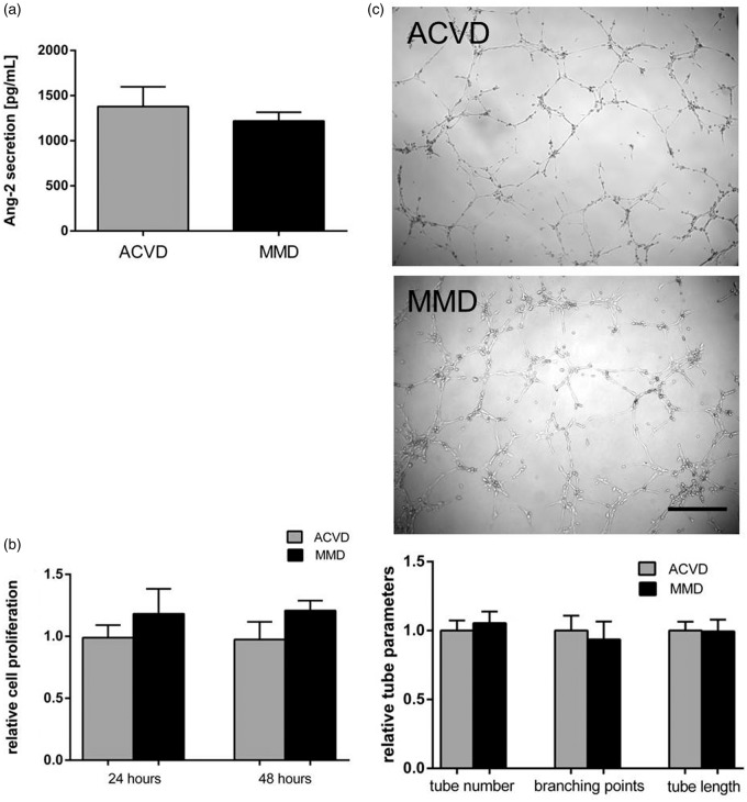
Figure 6.
ACVD versus MMD serum responses observed in non-cerebral ECs, HuVEC cells. HuVEC cells were incubated in MMD or ACVD serum for 48 h applying the experimental protocol used for cENDs. (a) Secretion of human Ang-2 into the supernatant was analyzed by ELISA. (b) HuVEC cell proliferation was not significantly changed in response to ACVD and MMD serum incubations, as measured by MTT assay. (c) Phenotypic effects of ACVD and MMD patients' serum incubation were tested by Matrigel tube formation assay. Representative low magnification images (10 × magnification) of tubule tube formation in response to ACVD and MMD serum were chosen. Total number of tubes, branching points, and tube length was assessed by Wimasis Image Analysis and depicted by the graph. Values are means ± SEM, (n = 3 independent experiments each including five different sera per group).

