Figure 1.
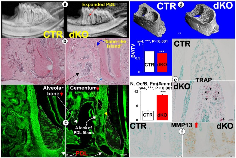
Eight-wk-old Bmp1/Tll1 double knockout (dKO) mice develop periodontal defects. (a) Representative X-ray images revealed alveolar bone loss and expanded periodontal ligament (PDL) in dKO mice (red arrow). (b) Hematoxylin and eosin staining showed alveolar bone loss (black arrow), a bone-like island (outlined in white), and much reduced cementum mass (black dashed line) with expanded PDL in dKO mice. (c) Confocal FITC images showed dKO alveolar bone loss, a lack of PDL fibers (yellow line), and reduced cementum mass with fewer cementocytes (white line). (d) Data from micro–computed tomography (µCT) analysis confirmed the dKO alveolar bone loss; quantitative analysis of bone volume (BV)/total volume (TV) of alveolar bone showed a statistically significant difference between dKO and control (CTR) samples (n = 4, ***P < 0.001). (e) Tartrate-resistant acid phosphatase (TRAP) stain images showed many more positive osteoclasts (black arrowheads) in dKO samples, which was statistically different from the control group (n = 4, ***P < 0.001), and (f) immunohistochemistry stains showed dramatic increases in matrix metallopeptidase 13 (MMP13) in dKO samples.

