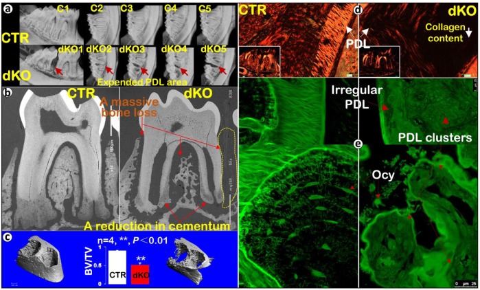
Figure 2.
The 17-wk-old Bmp1/Tll1 double knockout (dKO) mice display advanced periodontal defects. (a) X-ray images showed massive loss of alveolar bone between the dKO first and second molars. (b) Backscattered scanning electron microscopy images confirmed a great reduction in both bone and cementum masses in dKO mice. (c) Micro–computed tomography data showed advanced dKO alveolar bone loss, qualitatively and quantitatively (n = 4, **P < 0.01). (d) Sirius red staining imaged via polarized microscopy displayed an expanded dKO periodontal ligament (PDL) area with sharply reduced collagen fibers. (e) Images of fluorescein isothiocyanate staining displayed an irregularly organized dKO PDL layer with sparse, rounded osteocytes (Ocy). In contrast, in control samples, fibers filled in the entire PDL area and osteocytes were spindle shaped and well organized. BV, bone volume; CTR, control; TV, total volume.

