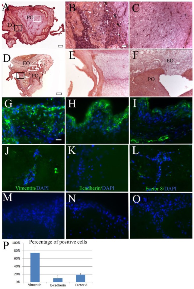
Figure 2.
Recell-dTBs after 1 wk in vitro bioreactor culture. (A) H&E-stained sectioned recell-dTBs. (B, C) High-magnification images of black-boxed EO (B) and white-boxed PO (C) areas in A. (D) H&E-stained acellular dTB construct. (E, F) Higher-magnification image of black-boxed EO (E) and white-boxed PO-EO border (F) areas in D. (G–L) IF staining was analyzed at the periphery (G–I) and centers (J–L) of all constructs for hDPCs expressing Vimentin (G, J), pDE cells expressing E-cadherin (H, K), and HUVECs expressing Factor VIII (I, L). (M–O) No positive staining was observed in negative controls. (P) Quantitative analyses showed that ~75% of cells were positive for Vimentin, 10% cells were positive for E-cadherin, and 19% cells were positive for Factor VIII. Scale bar = 2 mm in A and D; 20 µm in B, C, E, and F to O. DAPI, 4′,6-diamidino-2-phenylindole; dTB, decellularized tooth bud; EO, enamel organ; H&E, hematoxylin and eosin; hDPC, human dental pulp cell; HUVEC, human umbilical vein endothelial cell; IF, immunofluorescence; PO, pulp organ.

