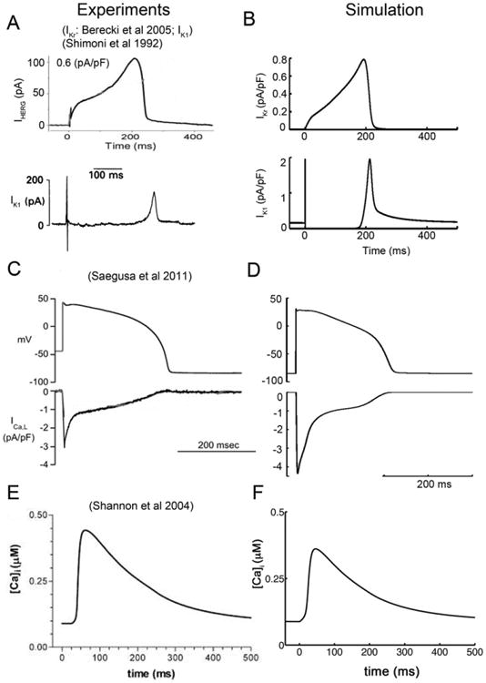
Fig. 1.

A comparison of experimentally recorded and model generated transmembrane ion currents from rabbit ventricular myocytes. (A) Experimentally measured IKr (upper) [55], and IK1 (lower) [14]. (B) Simulated IKr (upper) and IK1 (lower) compared. (C) Experimental action potential clamp waveform (upper) and corresponding L-type Ca2+ current (lower) from rabbit ventricular myocyte [56]. (D) Simulated rabbit ventricular myocyte action potential and model generated L-type Ca2+ current. (E) Experimentally recorded Ca2+ transient during the AP [15]. (F) Corresponding simulated Ca2+ transient.
